


Hypertemporal Imaging
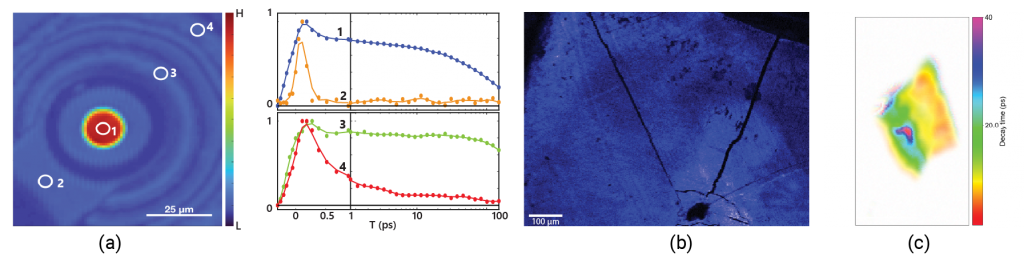
Figure 1. a) Lifetime of vertical cavity surface-emitting laser (VCSEL) diode; b) Charge transfer in perovskites; c) Defect lifetimes in compound semiconductors
With ultrafast temporal resolution, KRAKEN is able to measure ultrafast decay time maps. Compared to conventional ultrafast spectrometers, KRAKEN offers full images with transient absorption spectra measured at every pixel! These ultrafast decay maps provide insights into the inhomogeneity of material lifetimes and ultrafast dynamics.
Using KRAKEN users can also enhance contrast in an image using the ultrafast decay time. This is analogous to fluorescence/photoluminescence lifetime imaging microscopy (FLIM/PLIM), except KRAKEN is also sensitive to sub-picosecond timescales.
An example application: defects in many bulk semiconductors will trap electrons or excitons. Hypertemporal imaging distinguishes material regions based on the decay time of an excitation in the material. Long-lived trapped states isolated to defect sites offer KRAKEN users the ability to measure background-free signatures of defects.
Hyperspectral Imaging

Figure 1. a) Strain and composition in 2D materials; b) Defect states in microLEDs; c) Feature distinction in biological materials
Scanning the excitation wavelength using KRAKEN enables users to distinguish sample features based on the excitation wavelength. Hyperspectral imaging contrast arises from distinct material composition, strain, concentration, changes in electronic screening, Stark shifts, and more. Hyperspectral imaging reveals subtle changes in a material’s response that are easily missed with linear techniques.
Volumetric Imaging

Figure 1. a) 3D Morphology of semiconductor defects; b) Measuring layer structures in devices; c) Morphology of tissue
Ultrafast imaging is a nonlinear imaging technique, relying on the nonlinear interactions of pump and probe pulses in your material of interest. Signals due to nonlinear optical processes, like two-photon microscopy, primarily are emitted at the laser focus where the light is most intense. Ultrafast imaging is therefore naturally capable of volumetric imaging, with axial resolution that is comparable to two-photon microscopy.
When used in conjunction with hypertemporal or hyperspectral imaging contrast, KRAKEN can generate beautiful volumetric images of microscopy features in semiconductors, layered device structures, and tissue morphology.
Related Publications
- Joint white paper with Light Conversion featuring ultrafast microscopy for material characterization
- Joint white paper with Coherent featuring the applications of ultrafast microscopy to 2D materials
- Rapid ultrafast microscopy for characterizing chemical-vapor deposition (CVD)-grown materials
- Using silicon vacancy centers to probe strain tensor in diamond
- [FEATURED] Imaging dynamics in transition metal dichalcogenide heterostructures
- Pump-probe imaging microscopy to characterize historic artwork
- Pump-probe imaging microscopy to differentiate melanoma tissues
Broadband Operation
KRAKEN’s broadband tunable wavelength operation has automatic wavelength tuning, automatic focus control, and automatic alignment control. This enables rapid adjustment of wavelengths with the click of a button.
Minimal User Training
KRAKEN is designed to make measuring hypertemporal, hyperspectral, and volumetric ultrafast images accessible for a user-facility user on a tight schedule:
- Easy-to-use imaging interface
- Eye safe
- Non-contact, non-destructive
Stable Alignment
Collinear optical design made to maintain longterm alignment, which is unprecedented for ultrafast spectroscopy and imaging systems.
Active controls are also engineered to manage beam pointing change through extreme wavelength tuning from 450-1000 nm, along with normal beam-pointing fluctuations of the included ultrafast laser.
KRAKEN Multi-functional ultrafast spectrometer specifications
| Specification | Standard System | |
|---|---|---|
| Tunable wavelength range | 470-650 & 680-1000 nm | |
| Illumination power (at sample) | 0.5 – 10 mW | |
| Decay time range | 0-2000 ps | |
| Bandwidth-limited pulse duration | 120 fs | |
| Collinear Spectrometer | Transient Absorption Spectrometer *upgrade to BIGFOOT | |
| Compatible sample mounting | X-Y stage, Cryostat | |
| Spatial resolution | < 1 um | |
| Axial resolution | < 8 um (<3 um upgrade) | |
| Detection Image Frame Rate | 1 fps (20 fps upgrade) | |
| Dimensions | 62 in × 35 in × 65 in |
Try it out with a service inspection
Try out KRAKEN and ultrafast microscopy with our Advanced Material Characterization Services.
 Advanced Material Characterization Service
For hyperspectral, hypertemporal, and volumetric measurement of your advanced materials we offer a characterization service based on nonlinear imaging and spectroscopy.
This is a great way to test whether or not MONSTR Sense products would be a good fit for your lab, or to acquire some useful ultrafast imaging data for a paper or proposal.
For more information or to get on our mailing list please reach out to us at info@monstrsense.com or using our contact page.